Cell Division implantation Cell Division implantation Cell Division implantation
<Prev
logo
The Biology


The spinal column or backbone is made up of units of bone called vertebrae. These form in the early embryo from repeated cell groupings called somites. Depending on their position in the embryo the somites will form neck, thorax, lumbar or sacral vertebrae.


  Image. Repeating units of the developing backbone, somites, as they appear in the developing chick embryo. © Kate Storey

How it works

A series of somites are made sequentially along length of the embryo. The cells in each somite can tell which part of the backbone they are supposed to form based on when they were generated.


Mice have a ribcage with 12 pairs of ribs (left). When the gene Hox6 is activated in ribless regions of the embryo, extra ribs are formed, extending from the ribcage to the tail (right). Image: M. Mallo.


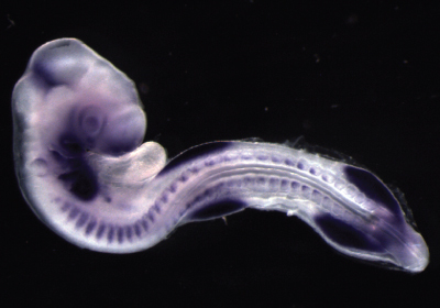
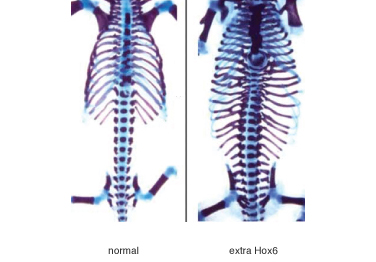
The somites that are made first are the oldest and make the bits of backbone in the neck. The cells do this in response to the activity of particular genes, known as Hox genes. The combination of Hox genes expressed changes as the later somites are added and so these acquire a different fate, becoming next thoracic vertebrae and so on. Somites are the first segmental structures to form in the embryo and the generation of these repeated units relies on a molecular clock, which is driven by oscillations of the Notch signalling pathway.

Find out more:
How Hox genes work

Key labs that work on development of vertebrae:

Andy Oates, NIMR/University College London
Olivier Pourquie, Harvard Stem Cell Institute
Denis Duboule, Ecole Polytechnique Federale de Lausanne
Jacqueline Deschamps
Moises Mallo, Instituto Gulbenkian de Cincia


Selected research papers:
A review of the segmentation clock, a molecular oscillator that regulates the periodicity of somite formation
Patterning embryos with oscillations: structure, function and dynamics of the vertebrate segmentation clock
The regulation of Hox gene expression during animal development


When development goes wrong


X-ray of patient with scoliosis

 
If the backbone is not made properly, the embryo can develop with a malformation called Scoliosis. This involves a curvature of the backbone and can be quite debilitating. The condition does not always have to be treated, but may be rectified with a plaster cast or a brace. A number of genes have now been identified that contribute to congenital and sporadic vertebral malformations, however, the causes of vertebral defects, like neural tube defects are likely to be multi-factorial and involve both genetic and environmental influences.


Find out more:
Scoliosis NHS information

Scoliosis Association UK – supports Scoliosis suffers in the UK

Selected research paper:
Review of progress in the understanding of the genetic causes of vertebral segmentation disorders in humans


Spinal column dress: Details


Spinal column dress: Silk chiffon (printed by Coats Viyella Plc), resin, aluminium leaf, fibre optic threads

The fabric patterning taken from a gene sequencing gel recalls the molecular basis of embryonic development. This female spine, cast in resin, was hand plated with aluminium foil. Fed through it are 8,000 fibre optic endings, representing the nerves extending out into the body.

Drawing
1997
Copyright Helen Storey


logoIntroduction / Biology & Design / Gallery / Film / Exhibition / Contact / Follow ProfHelenStorey on Twitter